RETeval® handheld ERG device
 We recently introduced new technology, the RETeval® handheld ERG (electroretinogram), into our practice to help us better diagnose and manage disease for our diabetic patients.
The RETeval device offers a functional test that gives us important information about the health of the retina. This objective assessment aids in the diagnosis of diabetic retinopathy and helps predict disease progression. By combining a retinal cell stress measure and a pupil light response, the device allows me to rapidly and noninvasively assess patients at risk for DR, as well as monitor retinal changes as the
disease progresses.
The results are delivered as a DR score, using a color-coded system that lets us know which patients are in the normal range and which are outside of normal limits. We are using this information to guide follow-up appointments, counsel patients, and more closely monitor those at risk of progression.
The test is comfortable for patients, uses 2 adhesive skin electrodes under the lower eye lids, requires no patient cooperation, and can be done in minutes.
We are excited to have this technology and believe it will have a positive impact on our mutual patients.
As we gather baseline data on patients, we will share that information and our treatment strategy with
you. Together, we can do more to intervene earlier in the disease process and enhance our patients’
vision and quality of life.
Corneal Mapping
Corneal topography, also known as photokeratoscopy or videokeratography, is a non-invasive medical imaging technique for mapping the surface curvature of the cornea, the outer structure of the eye. Since the cornea is normally responsible for some 70% of the eye's refractive power, its topography is of critical importance in determining the quality of vision.
The three-dimensional map is therefore a valuable aid to the examining ophthalmologist or optometrist and can assist in the diagnosis and treatment of a number of conditions; in planning refractive surgery such as LASIK and evaluation of its results; or in assessing the fit of contact lenses. A development of keratoscopy, corneal topography extends the measurement range from the four points a few millimeters apart that is offered by keratometry to a grid of thousands of points covering the entire cornea. The procedure is carried out in seconds and is completely painless.
Visual Field Testing
A visual field test measures how much 'side' vision you have. It is a straightforward test, painless, and does not involve eye drops. Essentially lights are flashed on, and you have to press a button whenever you see the light. Your head is kept still and you have to place your chin on a chin rest. The lights are bright or dim at different stages of the test. Some of the flashes are purely to check you are concentrating.
Each eye is tested separately and the entire test takes 15-45 minutes. Your optometrist may ask only for a driving licence visual field test, which takes 5-10 minutes. If you have just asked for a driving test or the clinic doctor advised you have one, you will be informed of the result by the clinic doctor, in writing, in a few weeks.
Normally the test is carried out by a computerised machine, called a Humphrey. Occasionally the manual test has to be used, a Goldman. For each test you have to look at a central point then press a buzzer each time you see the light.
AdaptDx Pro®
A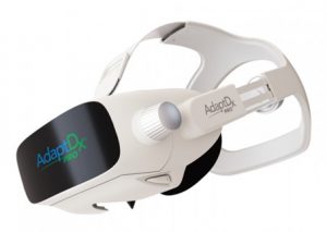 daptDx Pro leverages the science of dark adaptation testing to help detect age-related macular degeneration (AMD) up to three years before drusen are visible. A subclinical diagnosis gives you more time to slow the progression of AMD.
OPTOS Retinal Exam: Ultra-widefield Photography and Angiography
 Annual eye exams are vital to maintaining your vision and overall health. We offer the optomap® Retinal Exam as an important part of our eye exams. The optomap® Retinal Exam produces an image that is as unique as your fingerprint and provides us with a wide view to look at the health of your retina. The retina is the part of your eye that captures the image of what you are looking at, similar to film in a camera.
Many eye problems can develop without you knowing. You may not even notice any change in your sight. But, diseases such as macular degeneration, glaucoma, retinal tears or detachments, and other health problems such as diabetes and high blood pressure can be seen with a thorough exam of the retina.
Regular detailed examination of the inside of the eye – the retina, is critical to eye health. Doctors use a number of techniques to examine the retina including looking into the eye, usually after dilating and the use of special cameras for imaging inside the eye.
Until recently, most ophthalmic cameras could only photograph about 20% of the retina at a time. We now know that many eye diseases occur or begin at the outer edges of the retina, (“the periphery”), so examining this area is extremely important.
Because seeing the entire retina is so important at Ridgeview Optometry we have invested in the most advanced camera for ultra-widefield photography and angiography. In a single, quick shot, this camera produces “optomap” photos or angiograms of about 82% of the retina.
These optomap images provide superior visibility of the retinal periphery allowing us to see, document, show you, and follow pathology that could not be seen with traditional eye cameras.
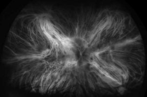


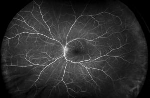
An optomap® Retinal Exam provides:
- A scan to show a healthy eye or detect disease.
- A view of the retina, giving your doctor a more detailed view than he/she can get by other means.
- The opportunity for you to view and discuss the optomap® image of your eye with your doctor at the time of your exam.
- A permanent record for your file, which allows us to view your images each year to look for changes.
The optomap® Retinal Exam is fast, easy, and comfortable for all ages. To have the exam, you simply look into the device one eye at a time and you will see a comfortable flash of light to let you know the image of your retina has been taken. The optomap® image is shown immediately on a computer screen so we can review it with you.
Please schedule your optomap® Retinal Exam today!
For more information on the optomap® Retinal Exam, go to the Optos website.

We are closed during the lunch hour, from 12:00 PM to 1:00 PM.